When Dan "threw out his back," the anatomical areas susceptible to injury align with the lumbar region of the spine, affecting the sciatic nerve and resulting in discomfort often referred to as the posterior area of the lower right limb. The imaging method employed for diagnosis would be Nuclear Magnetic Resonance Imaging.
Explanation:
Physical activities, such as lifting heavy items, can lead to lower back injuries which vary from painful muscle spasms, torn muscles, or harm to the lumbar spine's intervertebral discs.
In Dan's case, attempting to lift the furniture could lead to:
A herniated disc located between two vertebrae.
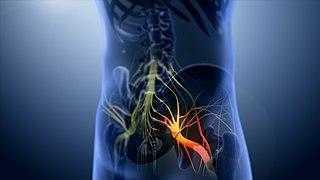
Herniated lumbar discs can put pressure on the sciatic nerve.
Damage to the sciatic nerve can cause a range of symptoms, including pain or nerve issues such as tingling, numbness, and sharp pains in the nerve's area.
Dan experienced pain in the posterior of his right leg where the compromised sciatic nerve travels.
Which imaging method would you suggest to identify a spinal issue?
The most suitable imaging technique to evaluate potential lesions for spinal injury diagnosis is Magnetic Resonance Imaging, which allows for the analysis of bone structure and soft tissues (muscle, cartilage) in the affected area.
X-rays only offer a glimpse of bone structures, aiding in the diagnosis of fractures, deformities, or structural changes, but do not reveal the actual condition of ligaments, intervertebral discs, or muscles.